Precancerous Lesions
The following lesions all have the potential to become malignant (lead to oral cancer).
Erythroplasia. Sometimes a red appearance may occur in the mouth due to unknown reasons. This is called erythroplasia. This is usually associated with smoking. The rate of transformation of erythroplasia to cancer is 5-10%.
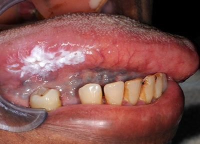
Leukoplakia is the term used to describe a white patch in the mouth that can not be rubbed off and can not be characterised clinically as any specific disease. Often these are related to smoking. The transformation rate in developed countries of leukoplakia to oral cancer has been estimated to be 2-6% in 10 years, although this may be higher in other parts of the world. The risk is greater when there are red areas associated with the white patch and when the lesion affects the tongue or floor of the mouth under the tongue.
Other lesions with the potential to become malignant include oral submucous fibrosis (which is associated with betel quid chewing and carries a high risk of malignant change), chronic candidosis (chronic fungal infection in the mouth), oral lichen planus (link to lichen planus page, oral discoid lupus and actinic keratosis (sun damage usually seen on the lower lip).
When a precancerous lesion is suspected then usually a biopsy will be suggested to exclude oral cancer and assess the risk associated with the lesion.
Treatment of these precancerous lesions will generally involve addressing the risk factors for oral cancer and monitoring the area. Sometimes removal of the lesion may be suggested.
Early oral cancers usually present as a white patch, a red patch, an ulcer, a lump or a raised area. Lip cancer usually appears as a crusty area with a raw surface. It is therefore important to have any new or changing lesions in your mouth checked by an oral healthcare professional.